A Quantitative Analysis Platform for PD-L1 Immunohistochemistry based on Point-level Supervision Model
Proceedings of the Twenty-Eighth International Joint Conference on Artificial Intelligence. Demos. Pages 6554-6556.
https://doi.org/10.24963/ijcai.2019/954
© 2019 International Joint Conferences on Artificial Intelligence
Keywords
- AI: Computer Vision
- Applications: Medical and healthcare
Abstract
Recently, deep learning has witnessed dramatic progress in the medical image analysis field. In the precise treatment of cancer immunotherapy, the quantitative analysis of PD-L1 immunohistochemistry is of great importance. It is quite common that pathologists manually quantify the cell nuclei. This process is very time-consuming and error-prone. In this paper, we describe the development of a platform for PD-L1 pathological image quantitative analysis using deep learning approaches. As point-level annotations can provide a rough estimate of the object locations and classifications, this platform adopts a point-level supervision model to classify, localize, and count the PD-L1 cells nuclei. Presently, this platform has achieved an accurate quantitative analysis of PD-L1 for two types of carcinoma, and it is deployed in one of the first-class hospitals in China.
- [Arteta et al., 2016] Carlos Arteta, Victor Lempitsky, J Al- ison Noble, and Andrew Zisserman. Detecting overlap- ping instances in microscopy images using extremal re- gion trees. Medical image analysis, 27:3-16, 2016.
- [Bearman et al., 2016] Amy Bearman, Olga Russakovsky, Vittorio Ferrari, and Li Fei-Fei. What's the point: Se- mantic segmentation with point supervision. In European conference on computer vision, pages 549-565. Springer, 2016.
- [Brahmer et al., 2012] Julie R Brahmer, Scott S Tykodi, Laura QM Chow, Wen-Jen Hwu, Suzanne L Topalian, Patrick Hwu, Charles G Drake, Luis H Camacho, John Kauh, Kunle Odunsi, et al. Safety and activity of anti-pd- l1 antibody in patients with advanced cancer. New England Journal of Medicine, 366(26):2455-2465, 2012.
- [Chen et al., 2017] Liang-Chieh Chen, George Papandreou, Florian Schroff, and Hartwig Adam. Rethinking atrous convolution for semantic image segmentation. arXiv preprint arXiv:1706.05587, 2017.
- [Dalal and Triggs, 2005] Navneet Dalal and Bill Triggs. His- tograms of oriented gradients for human detection. In international Conference on computer vision & Pattern Recognition (CVPR'05), volume 1, pages 886-893. IEEE Computer Society, 2005.
- [He et al., 2016] Kaiming He, Xiangyu Zhang, Shaoqing Ren, and Jian Sun. Deep residual learning for image recog- nition. In Proceedings of the IEEE conference on computer vision and pattern recognition, pages 770-778, 2016.
- [Hosang et al., 2017] Jan Hosang, Rodrigo Benenson, and Bernt Schiele. Learning non-maximum suppression. In Proceedings of the IEEE Conference on Computer Vision and Pattern Recognition, pages 4507-4515, 2017.
- [Jin et al., 2018] Qiangguo Jin, Zhaopeng Meng, Tuan D Pham, Qi Chen, Leyi Wei, and Ran Su. Dunet: A de- formable network for retinal vessel segmentation. arXiv preprint arXiv:1811.01206, 2018.
- [Laradji et al., 2018] Issam H Laradji, Negar Rostamzadeh, Pedro O Pinheiro, David Vazquez, and Mark Schmidt. Where are the blobs: Counting by localization with point supervision. In Proceedings of the European Conference on Computer Vision (ECCV), pages 547-562, 2018.
- [Li et al., 2018] Zeming Li, Chao Peng, Gang Yu, Xiangyu Zhang, Yangdong Deng, and Jian Sun. Detnet: A backbone network for object detection. arXiv preprint arXiv:1804.06215, 2018.
- [Long et al., 2015] Jonathan Long, Evan Shelhamer, and Trevor Darrell. Fully convolutional networks for seman- tic segmentation. In Proceedings of the IEEE conference on computer vision and pattern recognition, pages 3431- 3440, 2015.
- [Loy et al., 2013] Chen Change Loy, Ke Chen, Shaogang Gong, and Tao Xiang. Crowd counting and profiling: Methodology and evaluation. In Modeling, simulation and visual analysis of crowds, pages 347-382. Springer, 2013.
- [Reck et al., 2016] Martin Reck, Delvys Rodríguez-Abreu, Andrew G Robinson, Rina Hui, Tibor Csőszi, Andrea Fülöp, Maya Gottfried, Nir Peled, Ali Tafreshi, Sinead Cuffe, et al. Pembrolizumab versus chemotherapy for pd-l1-positive non-small-cell lung cancer. New England Journal of Medicine, 375(19):1823-1833, 2016.
- [Xue et al., 2016] Yao Xue, Nilanjan Ray, Judith Hugh, and Gilbert Bigras. Cell counting by regression using convo- lutional neural network. In European Conference on Com- puter Vision, pages 274-290. Springer, 2016.
- [Yu and Koltun, 2015] Fisher Yu and Vladlen Koltun. Multi- scale context aggregation by dilated convolutions. arXiv preprint arXiv:1511.07122, 2015.
References were extracted automatically from the PDF and may contain errors.
Full text extracted automatically from the PDF; figures, tables and equations may differ. The PDF is the version of record.
1 Introduction
Programmed death-ligand 1 (PD-L1) is known as a kind of proteins. The quantitative analysis of PD-L1 immunohistochemistry plays a very important role in the precise treatment of tumor therapy [Brahmer et al., 2012; Reck et al., 2016]. By quantitative analysis of the expression of PD-L1 in the tumor tissue of patients, it is beneficial to design a personalized medication strategy. The quantitative analysis of PDL1 requires classifying the cell nuclei into the positive tumor nuclei and negative tumor ones, as well as needing to accurately calculate the proportion of positive and negative ones respectively. Currently, in the process of quantitative analysis, pathologist (even in the first-class Hospital) manually calculate the cell nucleus. However, the sizes of whole slide PDL1 images ranges from several hundred megabytes to more than one gigabyte, in which the number of cell nuclei ranges from several thousands to even millions. Manual analysis not only takes a lot of time and effort, but is also extremely errorprone. Therefore, it is desirable to design an automatic and accurate quantitative analysis tool.
The quantitative analysis of PD-L1 does not require predicting the exact shape of the cell nucleus. As point-level annotations for the cell nucleus is relatively easy for pathologist to mark, and also can provide a rough estimate of the object locations, this kind of annotations is utilized to localize, classify and count the cell nuclei. Point supervision is widely adopted to object counting [Bearman et al., 2016; Laradji et al., 2018]. The solutions for object counting analysis can be classified into three categories: detection-based methods, regression-based methods, and density estimationbased methods [Loy et al., 2013]. Although the regressionbased methods, and density estimation-based methods are widely-used to predict the number of the detected objects, they cannot estimate the locations of the objects [Xue et al., 2016]. On the contrary, detection-based methods [Arteta et al., 2016] can be refined to precisely count the cell nuclei with their locations and classifications. In our platform, we choose the detection-based methods as the baseline.
With the point-level annotations of the cell nuclei, we adopt a fully convolutional networks (FCN) [Long et al., 2015] to quantitatively classify, localize, and count the PD-L1 cell nuclei. To reduce the counting error caused by cell nuclear stacking and deformation, HOG (histogram oriented gradients) [Dalal and Triggs, 2005] and NMS (Non-maximum suppression) [Hosang et al., 2017] are also applied to extract lowlevel features and filter negative positives. Using the aforementioned algorithms, we implement a platform for pathologist to automatically and quantitatively analyze the cell nuclei of PD-L1. Currently, this platform is used by pathologist in one of the first-class hospitals in China.
2 Implementation
2.1 The PD-L1 Annotation Tool
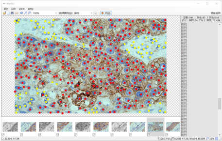
We have developed a PD-L1 annotation tool for pathologists, as shown in Figure 1. With this tool, the pathologist can quickly classify and locate the cell nuclei. The red dots represent the positive nuclei (i.e., tumor nuclei) and the yellow dots represent the negative nuclei (i.e., normal nuclei). In order to be able to make the analysis more accurate, we recommend that the pathologists mark the nuclei in the middle of its shapes. Empirically, it takes several minutes to finish a size of 1024 × 1204 images. The efficiency of the point-level samples collection is greatly improved compared to that of the mask-level samples collection.
2.2 Overview of the Platform
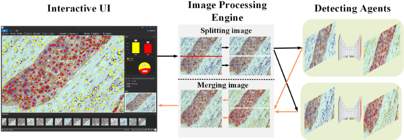
As shown in Figure 2, the platform mainly includes three parts: a front-end interactive UI, an image processing engine, and detecting agents.
- The interactive UI is primarily used to show the results of quantitative analysis, including the location, number, and proportion of negative and positive cell nuclei. Pathologists can directly browse the full-scale PD-L1 profiles of quantitative analysis results through the interactive UI.
- The image processing engine is mainly used to split the whole slide, distribute the divided sub-pictures, and merge the detecting results. In order to improve the efficiency of the inference, the processing engine splits the original PD-L1 image into several sub-pictures and uses the map-reduce computing framework to send those subpictures to multiple agents for the detection. Finally, the detecting results on different agents are combined together and sent to the interactive UI.
- The detecting agent contains a quantitative analysis model based on the FCN network [Long et al., 2015].
The backbone architecture uses a ResNet101 [He et al., 2016] network, and a dilation convolution kernel [Yu and Koltun, 2015] is also utilized to enhance the receptive field. We define an image-level loss to classify the nuclei into the negative ones and positive ones. Since the degree of nuclear staining, extrusion, and stacking affects the accuracy of counting and localizing, we use the HOG algorithm to calculate the nuclear split loss. Further, in the phase of the model inference, we adopt the NMS algorithm to filter the predicted point-level annotations to ensure that each cell nucleus contains only one annotation.
As the annotation tools provide an easy way for the pathologist to annotate pictures, thousands of PD-L1 pictures are collected for two kinds of carcinoma (i.e., lung squamous carcinoma and ductal breast carcinoma) in a short time. In the choice of detection network structure, we compare the performance between DUNet [Jin et al., 2018], DetNet [Li et al., 2018], DeepLab-v3 [Chen et al., 2017] and other models in detail. In our experiments, we find that the FCN network (with a ResNet101 as the backbone architecture) is more suitable for our scenario as the model has the highest accuracy and the lowest false positive rate.
3 Conclusion and FutureWork
Deep learning-based quantitative analysis of PD-L1 immunohistochemistry can not only greatly improve the diagnostic efficiency of pathologists, but also greatly improve the diagnostic accuracy. In this paper, we introduced a PD-L1 quantitative analysis platform based on the point supervision model. PD-L1 quantitative analysis is a good application of AI-based technology in the field of cancer therapy. Currently, we have achieved an accurate quantitative analysis of PD-L1 for two types of carcinoma. In the follow-up work, we will continue to expand the scopes of the platform among the hospitals.
Acknowledgments
This work was supported by the National Grand R&D Plan (Grant No. 2016YFB1000101).
@inproceedings{ijcai2019p954,
  title     = {A Quantitative Analysis Platform for PD-L1 Immunohistochemistry based on Point-level Supervision Model},
  author    = {Mi, Haibo and Xu, Kele and Xiang, Yang and He, Yulin and Feng, Dawei and Wang, Huaimin and Wu, Chun and Song, Yanming and Sun, Xiaolei},
  booktitle = {Proceedings of the Twenty-Eighth International Joint Conference on
               Artificial Intelligence, {IJCAI-19}},
  publisher = {International Joint Conferences on Artificial Intelligence Organization},
  editor    = {Sarit Kraus},
  pages     = {6554--6556},
  year      = {2019},
  month     = {8},
  note      = {Demos},
  doi       = {10.24963/ijcai.2019/954},
  url       = {https://doi.org/10.24963/ijcai.2019/954},
}
TY - CPAPER TI - A Quantitative Analysis Platform for PD-L1 Immunohistochemistry based on Point-level Supervision Model AU - Mi, Haibo AU - Xu, Kele AU - Xiang, Yang AU - He, Yulin AU - Feng, Dawei AU - Wang, Huaimin AU - Wu, Chun AU - Song, Yanming AU - Sun, Xiaolei T2 - Proceedings of the Twenty-Eighth International Joint Conference on Artificial Intelligence, IJCAI-19 PB - International Joint Conferences on Artificial Intelligence Organization PY - 2019 SP - 6554 EP - 6556 DO - 10.24963/ijcai.2019/954 UR - https://doi.org/10.24963/ijcai.2019/954 ER -Copied
Machine-readable versions of this paper, for tools and AI agents: