
Model Driven Visualization of Coronary Arteries
Proceedings of the Eighth International Joint Conference on Artificial Intelligence. Vision. Pages 1128-1130.
© 1983 International Joint Conferences on Artificial Intelligence
Abstract
In a joint project between the Departments of Computer and Information Sciences and Radiology we are applying techniques of artificial intelligence to improve clinical performance as related to coronary arteries. Specifically, we are investigating how images from intravenous digital subtraction angiography (DSA) can be enhanced so that their efficacy for lesion detection and quantitation becomes comparable with that of the more dangerous procedure of selective coronary arteriography. The enhancement techniques (which include algorithms for three-dimensional vessel detection, reconstruction, and display, as well as for accurate lumen size estjmation) are based on models of (i) the three-dimensional topological structure of the coronary arterial tree, (ii) myocardial dynamics, and (iii) the x-ray imaging process involved in producing digital subtraction angiograms. The evaluation of these model driven visualization techniques is done by the standard psychophysical method of Receiver Operating Characteristic (ROC) analysis applied to observer performance tests on images from an animal coronary atherosclerosis model.
- DeRouen, T. A., J.A. Murray, and W. Owen , "Variability of the Analysis of Coronary Artery Disease." Am. J. Cardiology 38 (1975) 174-178.
- Brody, W.R. (ed.), Conference on Ditigal Radiography, Proc. SP IE 314 (1981).
- Brown, B. G., E.L. Bolson, and H. T. Dodge, "Arte r iog r aphi c Assessment of Coronary Atherosclerosis," Arterios c l e r osis 2 (1982) 2-15.
- Ballard, D.H. and C.M. Brown, Computer Vision. Englewood Cliffs, NJ : Prentice-Hall, 1982.
- Bajcsy , R. and M. Tavakoli, "Computer Recog- nition of Roads from Satellite Pictures, " IEEE Trans. Syst. Man. Cyber . 6 (1976) 623- 637.
- Udupa, J.K., DISPLAY82 A System of Pro- grams for the Display of Three-Dimensional I nforma tion in CT Data. Medica l Image Pro- cessing Group Technic a l Report No. MIPG67. Department of Radiol ogy , Univ. of Penn- sylvania (1983).
- Herman, G.T., Image Re constru c tion from Pro- jec tions: The Fundamentals of Computerized Tomography. New York, NY : Academic Press, 1980.
- Brait, C., "Optimal Registration of Deformed Images," Ph.D. Dissertation, University of Pennsylvania, August 1981.
- Deutsch, E.S., "Thinning Algorithms on Rectangular, Hexagonal and Triangular Arrays," Com. ACM 15 (1972) 827-837
- Freeman, H., "Computer Processing of Line- Drawing Image~>," Comput . Surv. 6 (1974) 57- 97.
- Swets, J.A. and R.M. Pickett, Evaluation of Diagnostic Systems -Methods from Signal Detection Theory. New York, NY: Academic Press, 1982.
References were extracted automatically from the PDF and may contain errors.
Full text extracted automatically from the PDF; figures, tables and equations may differ. The PDF is the version of record.
MODEL DRIVEN VISUALIZATION OF CORONARY ARTERIES
Department of Radiology University of Pennsylvania 3400 Spruce Street Philadelphia, Pennsylvania 19104
ABSTRACT
In a joint project between the Departments of Computer and Information Sciences and Radiology we are applying techniques of artificial intelli gence to improve clinical performance as related to coronary arteries. Specifically, we are investigating how images from intravenous digital subtraction angiography (DSA) can be enhanced so that their efficacy for lesion detection and quantitation becomes comparable with that of the more dangerous procedure of selective coronary arteriography. The enhancement techniques (which include algorithms for three-dimensional vessel detection, reconstruction, and display, as well as for accurate lumen size estjmation) are based on models of (i) the three-dimensional topological structure of the coronary arterial tree, (ii) myocardial dynamics, and (iii) the x-ray imaging process involved in producing digital subtraction angiograms. The evaluation of these model driven visualization techniques is done by the standard psychophysical method of Receiver Operating Characteristic (ROC) analysis applied to observer performance tests on images from an animal coronary atherosclerosis model.
I THE MEDICAL PROBLEM
Conventional x-ray imaging uses photographic film or fluorescent material to produce an image of the x-ray shadow cast by the body. The intensity of the x-rays exiting the body along a given path through the body from the x-ray source to the detector will be dependent on the total absorption along the path. Structures that are much more absorbing of x-rays than the surround ing tissues will be easily identifiable in the x-ray images. However, the superposition of shadows wakes it very difficult to image struc tures that differ little from their surroundings, such as the coronary arteries .
Atherosclerotic coronary artery disease is a major public health problem. A noninvasive method of diagnosing coronary artery disease and
* The research reported in this paper is supported by National Institutes of Health grant HL29985.
*
Department of Computer and Information Sciences University of Pennsylvania Philadelphia, Pennsylvania 19104
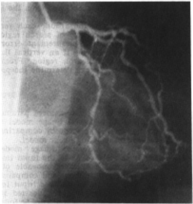
assessing the results of treatments, such as by pass graft surgery, would be a major contribution. Conventionally, coronary arteries are studied by selective arteriography, a somewhat dangerous procedure in which a catheter inserted into a specific artery is used to deliver contrast material to improve visualization of the lumen. See Fig. 1 for an example of a selective arterio gram. It has been asserted. that coronary arterio graphy has limitations in diagnosing vascular dis ease (see, e.g., [1]).
Vig~a£ ~ub~action angiog~aphy (DSA) is an alternative and much less i.nvasive test (see, e.g., [2]). X-ray images are obtained prior and after introduction of contrast into the circulatory system. The x-ray images are electronically detected and the detected x-ray intensities are digitized as functions of position. By knowing the initial x-ray intensity without contrast, the summed x-ray absorption of the contrast material alone can be calculated. If the patients remain motionless, the resulting subtraction image will have the shadow of nothing but the contrast material, and so in principle a much lower propor tion of contrast to blood need to be used than in selective arteriography. One can possible do without delivering the contrast to a specific site, and thus eliminate the need for insertion of the catheter into a specific artery and allow a simple intravenous injection of contrast. The quantitative capabilities of DSA offer the possibility of finding the relative and even absolute cross-sectional lumen area of the coronary vessels without r equiring sharp edge definition. This could provide a more accurate method than conventional selective arteriograpny, which relies on detecting variations in the sil houettes of vessels . In particular, the visual representation of computer-produced absolute measures of cross-sectional areas is likely to be a more repeatable and reliable predictor of the hemodynamic impact and of the prognostic significance of coronary stenosi~ (narrowing) than the currently commonly used subjective visual estimate of "percent stenosis" [3]. On the other hand, the image quality in DSA is inferior to that obtained with selective arteriography . Particular 1 i_mitations are decreased spatial resolution, and problems in separating overlying contrast in cardiac chambers with intravenous in jections. Hence the DSA images by themselves do not contain information for visualization that is competitive with selective arteriography . On the other hand, a great deal of a priori knowledge exists (such as gross anatomy) and can be put into models which provide information for driving visualization procedures.
The problem that we are investigating is the following: "Can the application of model driven visualization procedures to images produced by DSA improve their clinical efficacy to or above the level of the efficacy of the more dangerous procedure of selective coronary arteriography?"
The phrase model dniven v~uafization is used here in a standard sense as defined, for example, by Ballard and Brown [ 4]: "high-level models in the knowledge base generate expectations as pre dictions of geometric, segment, or generalized image structures in the input".
II VISUALIZATION TECHNIQUES
In conventional coronary arteriography, pro jection images are obtained from several direc tions on high contrast film. The outline of the vessels is studied and the severity of local vascular disease is assessed by the degree of narrowing of the outline. As atherosclerotic lesions are commonly eccentric and irregular, several projections are made, and the one showing the greatest degree of narrowing for a given lesion is used to estimate the severity of the lesion. The size of the lesion often tends to be underestimated from the images, either because the lesion was not imaged tangentially to its dimension of greatest narrowing or because the local narrowing was part of a longer segment of more diffuse disease. On the other hand, post stenotic dilation could also lead to overestima tion of degree of stenosis. See e.g., [3] for a discussion.
In DSA, a bolus of contrast medium is inject ed intravenously and sequential digitized fluoro scopic images are obtained as the bolus of con trast medium flows through the central circulation. To improve the images, initial images, without con trast medium present (masks), are digitally sub tracted from the later images.
Two major difficulties with DSA are that (i) the vessels overlay the beating heart, with con trast material in the chambers superimposed on the vessels, and (ii) images are less sharp than those obtained with selective arteriography. A poten tial advantage of DSA is that the quantitative nature of the image data may allow direct calcu lation of the relative cross-sectional areas of the vessel lumens from the integrated profile of the vessel image. This would allow detection of regional vascular disease without requiring sharp edge definition, and might even permit more accu rate assessment of severity of lesions than con ventional arteriography . In order to realise this potential, very accurate digital masking must be performed, including: nonlinear (logarithmic) subtraction, correction of possible effects from x-ray beam hardening by dense absorbers, such as bones, EKG gated imaging to include the effects of cardiac motion, and a model for the contrast in the cardiac chambers. The hierarchical masking procedure which we now describe is based on models of both the myocardial dynamics and the x-ray imaging process involved in DSA.
Our time variant subtraction mask for the digital imaging is built from three components. The first component of the mask includes the time invariant structures, such as bones and soft tissues of the chest wall and abdomen (the studies are performed during suspended respiration to avoid respiratory motion). The second component of the mask incorporates the moving portion of the image before the injection of contrast, as a func tion of phase of the cardiac cycle (determined by EKG gating). The third component of the mask includes the non-cyclic effects of the cardiac chambers (for studies made with intravenous injec tions). These are modelled as four chambers with cyclical dimension changes, each with an average contrast concentration that changes smoothly with time but is uniform within each separate chamber at any particular time.
The production and subtraction of masks pro vide us with images which are supposed to be pro jection images of the three-dimensional vessel structure only. Since this can only be achieved approximately, two-dimensional image processing applied to these images can improve their efficacy. In addition to standard image enhancement techni ques, we are investigating the usefulness of a model driven detection system which makes use of built-in knowledge of properties of the appearance of objects it is supposed to detect. We are adopt ing the general approach of [5], which reports on a model driven approach for finding roads in satellite images, but use of course a different model which incorporates the expected appearance of vessels in DSA images. Such image enhancement techniques are needed, since in the unenhanced images the "noise" is likely to be high enough to cause local areas in coronary arteries to be below the threshold of observer delectability. Model driven two-dimensional enhancement can be used to produce new (high contrast) images.
To implement the further processing of the images, to create new images incorporating informa tion on the cross- sectional areas of the vessel lumens , several steps are necessary. First, the 3dimensional orientation of the vessels must be determined from the projection images . While this would not be possible for a given general set of limited projection images, we can use our know ledge of the special properties of the vessels to help us, including the connectedne ss of the vessels, the limitations on numbers and angles of branches , and the fairly standard arrangement of the larger vessels.
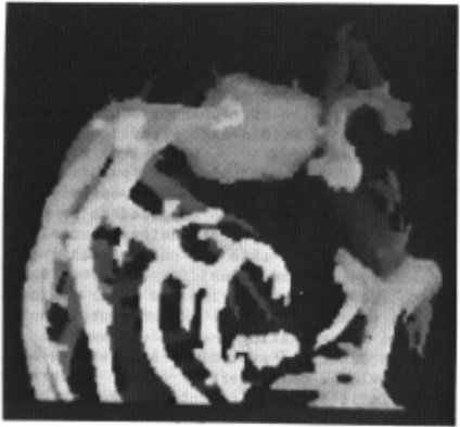
Thinning algorithms that we have developed for the analysis of carotid arteriograms are used to determine the local orientations in 3-dimens ions of the lumens of the vessels . The integrated area beneath the profiles of the vessels at right angles to the direction of the axis is determined in the projections and the results are correlated with the 3- dimensional orientations of the vessels to produce a map of estimated vessel lumen cross sectional area along the vessels. This informa tion can be displayed in several ways . The area can be encoded on either a gray scale intensity or a pseudo color scale and used to modify the appear ance of the vessel in the display. Area can be displayed relative either to an overall scale or to the segments of vessels between branch points . The area information can be used as an overlay on the original images of the vessels . It can also be used in conjunction wi t h the 3-dimensional map of the vessels to create an interactively rotatable 3-dimensional display of the vessels , as illustrat ed in Fig. 2, see [6 ] for details.
i~e now briefly discuss three of the techniques that are used to achieve the aims outlined above .
Three-dimensional reconstruction from multiple projections is now a well- developed field [7]. It is however well known that accurate rec'onstruction from a few projections is not possible unless a great deal of a priori knowledge is available . In the project under discussion we make use of a priori knowledge based on a model of the topo logical structure of the coronary arterial tree, as well as the knowledge that arteries lie on the surface of the myocardium.
Both in the 3D reconstruction (where a model of the tree has to be matched with the coronary tree of a specific individual) and in the modell ing of myocardial dynamics (for the production of subtraction masks) we make use of an elastic deformation method, in which we map a reference object onto an actual object under the assumption that the reference object is made from an elastic material which is then deformed to become similar to the actual object [8] . The mapping of choi ce is one which minimizes a cost function of the form "DEFORMATION minus SIMILARITY".
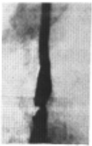
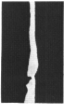
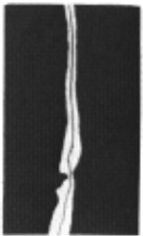
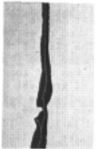
We complete this section with an example s how ing why we need model driven variants of general algorithms . For accurate "lumen cross- sectional area" estimation , we need to define the " direc tion" of the vessel. This we do by using a medial line to determine the direction. In Fig. 3a we show a DSA image of a common carotid artery with a very noticeable s t enois . To find the medial line we first applied thresholding to produce Fig. 3b. Our model driven adaptation of the parallel thining algorithm reported in [9] produced Fig. 3c which shows an acceptable representation of the direction of the vessel all along its length. The unalter ed algorithm of [9] could not distinguish the stenosis from a branch (since it had no know ledge of such things) and produced the i nappropri ate (for our application) medial line shown in Fig . 3d .
'"'
(h)
(c! \
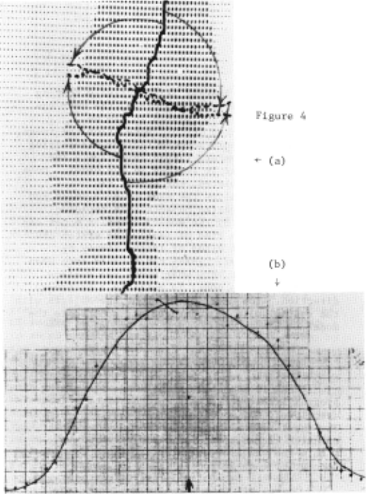
The medial line so ob t ained can be chaincoded [10] , and thus easily rotated by 90° in both di rections at any point along i t s length (see Fig . 4a) , defining two "normals " t o the direction a t tha t point . The i ntensi t y values in the original (Fig. 3a) at points corresponding t o points on these normals can be plotted (Fig . 4b), and the a rea under this cur ve can be used as a measure of the lumen cross-sectional area . ·················· ..... ................
III EVALUATION
We apply our model driven enhancement techni ques to images obtained by using a clinical DSA unit on anima ls with coronary artery lesions. The usefulness of the techniques is tested with a Receiver Operating Charac teric (ROC) analysis [11]. Th-e perfo rmance of observers in detecting lesions in the original DSA images is compared with their success with the enhanced images and with con ventional selective coronary a rteriography, con sidering both increased sensitivity and possible increased false positive diagnosis. The truth or falseness of detected lesions is judged by compar ing t o specimen angiograms. To increase the number of images for testing, additional synthetic lesions of different size and location are created in the digital images and the ability to detect them with diffe r ent t ypes of enhancement is studied.
IV DISCUSSION
We have described work which is currently be ing pursued as a coope r ative project be tween the Department of Computer and Information Sciences and Department of Radiology a t the University of Pennsylvania . The projec t involves applying techniques of artificial intel ligence to sol ve a significant medical problem. It i s our conjec ture that the model driven visualiza t ion procedures described above wil l make DSA at leas t as useful as the more dangerous currently used procedure of selec t ive arteriography. At the time of wri ting, many of the above-described visualization proce dures have been implemented, but the evalua tion on the animal model has only jus t s t ar t ed . Hence validation (or refutati on) of our conjecture can not as ye t be given.
ACKNOWLEDGEMENTS
The author s are grateful t o R. Summe r s , G. Waxle r, and M. Wiener for programming help, S. Strommer and P. Porter fo r the phot ography , and M.A. Blue for the t yping .
@inproceedings{ijcai1983p262,
  title     = {Model Driven Visualization of Coronary Arteries},
  author    = {Herman, Gabor T. and Axel, Leon and Bajcsy, Ruzena and Kundel, Harold L. and LeVeen, R. and Udupa, Jayaram K. and Wolf, G.},
  booktitle = {Proceedings of the Eighth International Joint Conference on
               Artificial Intelligence, {IJCAI-83}},
  publisher = {International Joint Conferences on Artificial Intelligence},
  editor    = {Alan Bundy},
  pages     = {1128--1130},
  year      = {1983},
  note      = {Vision},
  url       = {https://ijcai.org/proceedings/1983/262},
}
TY - CPAPER TI - Model Driven Visualization of Coronary Arteries AU - Herman, Gabor T. AU - Axel, Leon AU - Bajcsy, Ruzena AU - Kundel, Harold L. AU - LeVeen, R. AU - Udupa, Jayaram K. AU - Wolf, G. T2 - Proceedings of the Eighth International Joint Conference on Artificial Intelligence, IJCAI-83 PB - International Joint Conferences on Artificial Intelligence PY - 1983 SP - 1128 EP - 1130 UR - https://ijcai.org/proceedings/1983/262 ER -Copied
Machine-readable versions of this paper, for tools and AI agents:
 Welcome
 Future Conferences
 AI Journal Division
 IJCAI Proceedings
 About IJCAI
 IJCAI Awards