Automatic Rib Detection in Chest Radiographs
Proceedings of the Fifth International Joint Conference on Artificial Intelligence. Vision. Pages 697-697.
© 1977 International Joint Conferences on Artificial Intelligence
Abstract
Full text extracted automatically from the PDF; figures, tables and equations may differ. The PDF is the version of record.
AUTOMATIC RIB DETECTION IN CHEST RADIOGRAPHS
C. M. Brace(1), J. H. Kulick(1) "". W. Challis(2) (1) Department of Computing and information Science (2) Department of Diagnostic Radiology Queen's University Kingston, Ontario CANADA
ABSTRACT
This paper presents an overview of an algo rithm for rib detection in chest x-rays. The goal of the algorithm is to return the location of the borders of the ribs to a screening system to en able intercostal spaces to be extracted and analysed.
The detection of objects in pictures can usu ally be separated into two steps: image enhance ment and feature extraction. The amount of compu tation required by our algorithm is minimized by processing only that information which is required to achieve the goal of the algorithm, and by using knowledge of the structure of the rib cage to guide the extraction of the rib borders. In par ticular, significant reduction in computation is achieved by avoiding the enhancement of the entire picture. In addition, not all points in the image need be considered as possible rib border points, nor do all points on each rib border need be detected.
The rib borders are returned to the screening system as second degree polynomials. Results of testing the algorithm for a set of chest radio graphs are illustrated by superimposing the detected rib borders onto the X-ray images.
l-l Previous Work
The existing algorithms for automatic rib detection by Toriwaki (Tor-73A) Wechsler (Wec-75A, Wec-758) and Persoon (Per-75~ have all taken the approach of first preprocessing the en tire image by global operations to enhance it and then finding all the points on each rib border.
If these algorithms are considered carefully, it becomes apparent that much of the computation done by these three methods is not necessary. In all three algorithms, every point on each rib border is, at sometimei located. As each border is represented b~ a ow order polynomial, only a small number of po1nts need be located on each curve. Furthermore, performing an operation on the whole picture or considering all points as equal candidates for rib borders is both unneces sary and expensive.
Z-l Overview of the Algorithm
Detection of the rib borders precedes separately for each lung. The initial step lo cates the lower border of two adjacent ribs in the upper middle portion of the lung field; the aver age width of one of these starting ribs is also calculated. The next step selects vertical slices through the lung field, enchances them, and detects rib border points. The location of the two starting ribs and the average rib width is used to guide the search on each slice for the remaining ribs. When border points have been detected on all slices~ they are linked together to form a set of points ror each rib border. Fi nally, second degree polynomials are fitted to each border.
Z-Z Detection of the Starting Ribs
In order to determine candidate areas for the
This work has been supported by Medical Research Council of Canada grant MA5614 1 and by National Research Council of Canada grant A9279.
detection of all the rib borders on each slice, two quantities are required: 1) the distance between two adjacent ribs and 2) the average width of one of these ribs.
An extensive search for the starting ribs is necessary because 1) the starting ribs must be ac curately found in order to provide information for the selection of candidate areas for the remaining ribs, and 2) there is no prior knowledge about their expected positions or width to guide the search. The detection of the starting ribs proceeds in the following 5 steps:
jJ The Area of Search ~ Selected
A section of the image is chosen which lies midway in the lung field between the bottom and the top of the lung. The section is 128 columns wide and 64 rows high (the original image is 256x256)j this covers an area of the image from the med1astinum to the outside of the thorax and is large enough to include at least two complete rib images.
ii) The Selected Area Is Pre-processed
The selected region is pre-processed using hysteresis smoothing (Dud-74). The smoothing win dow height is chosen dynamically via an analysis of a grey level distance histogram.
iii) Detection of Possible Rib Borders
A funcion, MRIB, is applied to each of the smoothed columns. MRIB measures the amount a given interval on a slice corresponds to a rib im age, by comparing the average grey level within an interval with that outside the interval. MRIB is applied using a range of potential starting points and rib widths. Essentially MRIB behaves like a correlation function.
iv) Linking Two Sets of Border Points
The number of possible border points occur ring at each of the 64 rows of the section is cal culated. Two of the rows, R1 and R2, are selected as the rows containing the largest number of pos sible border points provided that these rows are between 10 and 30 rows apart. R1 and R2 are as sumed to occur at the peaks in the curves of two adjacent ribs.
Two sets of points are formed as the lower border points of the two ribs. The first points entered in each sets are those possible border points in smoothed columns which are closest to R1 and R2, respectively. The remaining points in the sets are selected from the possible border points in columns that are closest to the previously selected points.
y) Calculation of the Average Rib Width
The set of most likely widths of ribs is avera~ed to obtain the width of one of the start ing r1bs.
Z-1 Detection of Other Ribs
The other ribs are detected by searching above or below already known rib borders in an area in which the next rib is expected to be lo cated. Relationships between inter-rib spacings have been demonstrated by Persoon (Per-75), and verified by us. The rib border points are found on a number of vertical slices through the lung fields. The slices are spaced several columns apart as not all points on the rib border are necessary.
3-l Experiments
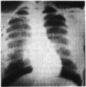
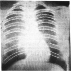
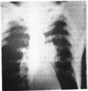
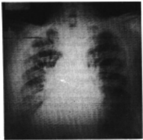
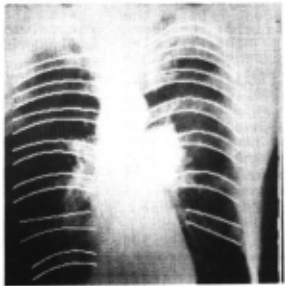
Our rib detection algorithm has been used to detect ribs in several chest radio~raphs. These X-rays show a variety of qualit1es, both in disease state and in film quality. ~o illustrate the results of the algorithm, the detected borders have been superimposed onto the image. Figures 1a-1c show each radio~raph; Figures 2a-2c show the same x-ray with the r1b borders superimposed.
As can be seen in the illustrations, the al gorithm is fairly successful for good quality films; usually 7 pairs of ribs are detected. Failure to detect a rib has five main causes:
- The clavicle. Because the image of the clavicle is similar in nature to the rib images and crosses the ribs at the top of the lung, rib border points are sometimes chosen which are actu ally borders of the clavicle. ~his occurs more frequently in images with very dark lung fields, such as in the image shown in Figure 1-c, where the clavicle is more visible than the ribs.
- ~he diaphragm area. If the bottom rib is close to the diaphragm, or the diaphragm does not have a sharp border (Figure 1-c), points may be detected in the diaphragm instead of at the ribs . or the points may be missed. In add it ion, if breast tissue is present in the X-ray then points may be detected in that tissue near the outside of the thorax rather than on the rib below.
- ~he heart border. ~he actual edges of the heart may be sufficiently lower in gray level than the estimated white value to allow some border points of the heart to be detected as rib borders. !hese points usually lie close enough to the actu al ribs to be included as border points but far 8nough away to make the curves representing the ribs inaccurate.
- Dark areas. If some of the ribs are not distinguishable in very dark lung fields, then several border points may be missing from those ribs. If there are large gaps in the · ribs then the sets of border points may be incorrectly linked and the resulting curves may be inaccurate or unacceptable. ~his is the cause of missing rib borders in Figures 1-b and 1-c.
5) Disease. If disease obscures the ribs then border points may be missing or inaccurate. ~he algorithm is able to overcome tumours which do not completely obscure the ribs but performs poor ly when a large amount of disease is present as in Figure 1-c. In such cases the ribs are not visi ble anyway and the X-ray should be considered as abnormal.
BIBLIOGRAPHY
Dud-73 Duda,R.O. and P.E. Hart, Pattern Classification and Scene Analysis, New York: John Wiley, 1973.
Per-75 - Persoon,E. "A New Edge Detection Algo rithm and its Application in Picture Processing", School of Electrical Engineering, Purdue Universi ty, TR-EE 75-38, October, 1975.
Tor -73A- Toriwaki,J., Y.Suenaga, T.Negoro, T.Fukumura, "Pattern Recognition of Chest X-Ray Images" 1 Com~uter Graphics and Image Processing 2, pp.252-~71' 973. .
Wec-75A- Wechsler,H. and J.Sklansky, "Automatic Detection of Contours of Ribs in Chest Radio ~raphs", School of Engineering, University of Cal lfornia, Irvine, TR-75- 1, February 1975.
Wec-75B- Wechsler,H., "Automatic Detection of Rib Contours in Chest Radiographs", PhD Dissertation, University of California, Irvine, 1975.
Vis i on-8: Br<lcP. 698
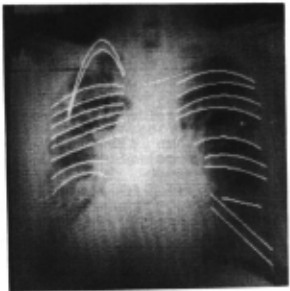
Figure 2-c
@inproceedings{ijcai1977p165,
  title     = {Automatic Rib Detection in Chest Radiographs},
  author    = {Brace, C. M. and Kulick, J. H. and Challis, T. W.},
  booktitle = {Proceedings of the Fifth International Joint Conference on
               Artificial Intelligence, {IJCAI-77}},
  publisher = {International Joint Conferences on Artificial Intelligence},
  editor    = {Raj Reddy},
  pages     = {697--697},
  year      = {1977},
  note      = {Vision},
  url       = {https://ijcai.org/proceedings/1977/165},
}
TY - CPAPER TI - Automatic Rib Detection in Chest Radiographs AU - Brace, C. M. AU - Kulick, J. H. AU - Challis, T. W. T2 - Proceedings of the Fifth International Joint Conference on Artificial Intelligence, IJCAI-77 PB - International Joint Conferences on Artificial Intelligence PY - 1977 SP - 697 EP - 697 UR - https://ijcai.org/proceedings/1977/165 ER -Copied
Machine-readable versions of this paper, for tools and AI agents: